Many myths and misconceptions have dominated glaucoma treatment for the past half century. At this year’s World Glaucoma Congress in Paris, Felipe Medeiros, MD, PhD, of the University of California, San Diego, highlighted several pertaining to IOP. First and foremost, he cited the idea that an IOP of 21 mm Hg is what separates healthy patients from glaucoma patients. The number 21 came about in population studies during the 1950s but in no way indicates the presence or absence of glaucoma. Second, he proposed that the nomograms used to correlate central corneal thickness and IOP are invalid and should therefore be used with caution in planning glaucoma care. Finally, he dissolved the myth that IOP is highest in the morning. In actuality, IOP is highest during sleep, when patients are lying down. Supine-position tonometry may be more relevant than diurnal curves.
A growing awareness of misconceptions like these have led to ophthalmologists’ use of a variety of analytical devices to aid their detection of glaucoma and its progression. No physician treating glaucoma patients can do so without the assistance of an optic nerve analytical device such as the HRT (Heidelberg Engineering GmbH, Heidelberg, Germany), GDx (Carl Zeiss Meditec, Inc., Dublin, CA), or Stratus OCT (Carl Zeiss Meditec, Inc.) in combination with advanced visual field perimetry.
Over the past decade, the development of minimally invasive glaucoma surgeries (MIGS) has lowered ophthalmologists’ thresholds for the surgical treatment of glaucoma. MIGS procedures reduce surgical time and complications and are cosmetically more acceptable. They allow for a more rapid visual recovery than the gold standards of trabeculectomy ab externo or large aqueous drainage devices like the Baerveldt glaucoma implant (Abbott Medical Optics Inc., Santa Ana, CA), Molteno Implant (Molteno Ophthalmic Limited, Dunedin, New Zealand), or the Ahmed Glaucoma Valve (New World Medical, Inc., Rancho Cucamonga, CA). After performing several MIGS procedures in my clinical practice, I will caution surgeons that these new devices and techniques are not one size fits all. They should be aware of the limitations of each prior to planning for surgery. This month’s “Peer Review” column highlights articles on this subject published during the past several years.
The following MIGS devices and procedures have gained popularity:
- Ex-Press Glaucoma Filtration Device (Alcon Laboratories, Inc., Fort Worth, TX):
This 0.4- X 3-mm stainless steel minishunt drains aqueous fluid into the subconjunctival space and is used in conjunction with standard trabeculectomy surgery. - Canaloplasty:
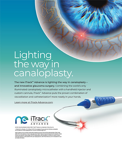
Canaloplasty is performed by passing a 9–0 or 10–0 Prolene suture (Ethicon, Inc., Somerville, NJ) through 360º of Schlemm canal with the aid of an iTrack (iScience Interventional, Menlo Park, CA) microcatheter and viscoelastic to dilate the canal. The procedure requires the creation of a deep sclerectomy with a flap and no antimetabolites to reduce the IOP without the formation of a bleb. - iStent (Glaukos Corporation, Laguna Hills, CA; not available in the United States):
This small, transtrabecular, titanium stent drains aqueous fluid into Schlemm canal. The device is placed through a clear corneal incision with the aid of a gonioscopy lens. The iStent does not aggressively lower the IOP and should be reserved for mild-to-moderate cases of glaucoma. - CyPass Micro-Stent (Transcend Medical, Menlo Park, CA; not available in the United States):
This supraciliary microstent is implanted through a clear corneal incision and is designed to increase uveoscleral outflow. - Solx Gold Shunt (Solx Inc., Waltham, MA; not available in the United States):
The Solx Gold Shunt is a 3- X 6-mm device that is less than 0.1 mm thick. It is placed into the supraciliary space through a 3-mm incision to increase uveoscleral outflow. - Endoscopic Cyclophotocoagulation (ECP):
ECP selectively ablates the pigmented ciliary epithelium to reduce aqueous production. The procedure is performed under direct visualization with a 20-gauge curved probe (E2 Microprobe Laser and Endoscopy System; Endo Optiks, Little Silver, NJ), which consists of a fiber optic camera and a diode laser that emits pulsed, continuous-wave energy at 810 nm. The ECP probe controls the precise placement of the laser energy. ECP is typically used to treat 360º of the ciliary processes. - Trabectome (NeoMedix Corporation, Tustin, CA):
This thermal cautery device ablates a 2- to 4-clock-hour segment of trabecular meshwork and Schlemm canal under direct visualization with a gonioscopy lens.
I hope you enjoy this installment of “Peer Review,” and I encourage you to seek out and review the articles in their entirety at your convenience.
LASER TRABECULOPLASTY
In a retrospective, noncomparative study, researchers found that previous laser trabeculoplasty did not appear to significantly affect the IOP but might increase the need for glaucoma medications in patients undergoing ab interno surgery using the Trabectome. From the Trabectome Study Group’s patient database, researchers analyzed outcome measures from 1,345 patients with open-angle glaucoma (OAG), including IOP, number of glaucoma medications, and the occurrence of secondary procedures. In eyes that had previously undergone trabeculoplasty, the mean IOP was 16.5 ±4.0 mm Hg, with an average decrease of 24% from the preoperative IOP at 1 year. In eyes that had not undergone previous trabeculoplasty, the mean IOP measured 15.7 ±3.0 mm Hg, with an average decrease of 30% from the preoperative IOP at 1 year. The use of adjunctive medications decreased to 2.1 and 1.5, respectively, in eyes that had and had not previously undergone trabeculoplasty.1
STENT IMPLANTATION
iStent
In a prospective, clinical study, 33 patients with OAG or ocular hypertension were randomized to one of two treatment groups. Group 1 received two iStents and underwent cataract surgery, whereas group 2 underwent cataract surgery alone. Postoperatively, aqueous flow and trabecular outflow were similar in group 1 (1.78 ±0.44 mm Hg and 1.74 ±0.82 mm Hg; P = .18) and group 2 (0.12 ±0.03 mm Hg and 0.13 ±0.06 mm Hg; P = .71). After surgery, there were no changes of note in aqueous flow, but trabecular outflow increased in both groups. At 1 year, trabecular outflow had increased by 275% in group 1 and 46% in group 2. The mean reduction in IOP was also greater in group 1 than in group 2 (6.6 ±3.0 mm Hg vs 3.9 ±2.7 mm Hg; P = .002). The mean number of medications used postoperatively was significantly lower in group 1 than in group 2 (0.0 vs 0.7 ±1.0, respectively; P = .007).2
In a prospective, randomized, open-label, controlled, multicenter clinical trial, 240 eyes with mild-to-moderate OAG were randomized to undergo cataract surgery with the implantation of the iStent or cataract surgery alone. The IOP of the selected eyes was controlled (≤ 24 mm Hg) on one to three medications. The primary measurement of effectiveness was an IOP of less than 21 mm Hg at 1 year with no medications. The secondary measurement was an unmedicated reduction of IOP by more than 20% at 1 year. Seventy-two percent of the combined group and 50% of the surgery-alone group achieved the primary outcome (P < .001). One year postoperatively, the IOP in both treatment groups was statistically significantly lower than baseline values. Sixty-six percent of the combined group and 48% of the surgery-only group achieved more than a 20% reduction in IOP without medication (P = .003). The overall incidence of adverse events was similar between groups with no unanticipated adverse effects.3
In a prospective, double-masked, randomized clinical trial, 33 patients with primary OAG were scheduled to have phacoemulsification with the implantation of an IOL (control group) or phacoemulsification with the implantation of an IOL and the insertion of an iStent (combined group). The mean baseline IOP was 17.3 ±3.0 mm Hg in the control group and 17.9 ±2.6 mm Hg in the combined group (P = .512). The mean IOP was 15.7 ±1.1 mm Hg in the control group and 14.8 ±1.2 mm Hg in the combined group at 15 months. It was 19.2 ±3.5 mm Hg and 16.6 ±3.1 mm Hg, respectively, 16 months postoperatively, following a 1-month washout of ocular hypotensive agents. The IOP was statistically significantly higher in the control group than in the combined group at both time points (P = .042 and P = .031, respectively). At 15 months, the mean number of medications used was lower in the combined group than in the control group (0.4 ±0.7 and 1.3 ±1.0, respectively; P = .007).4
Ex-Press Glaucoma Filtration Device
In a retrospective, consecutive, case-controlled study, data were collected from the charts of 35 patients who received the Ex-Press Glaucoma Filtration Device and 35 patients who underwent trabeculectomy. At least 2 years of follow-up data were available for all patients. IOP, bleb morphologic features, reduction of dependence on medication, visual recovery, number of postoperative visits, and postoperative complication rates were compared between the two groups. Mean IOP measurements were similar after 6 months but became slightly higher in the Ex-Press group at 1 year and at the final follow-up visit (P = .004 and P = .008, respectively). An unqualified success was defined as an IOP of between 5 and 18 mm Hg and at least a 30% decrease in IOP without IOP-lowering medication. A qualified success was the same as an unqualified one but with the use of IOP-lowering medications. An unqualified success was achieved with 77.14% of Ex-Press and 74.29% of trabeculectomy procedures at the final follow-up visit (P = 1.00). An additional 5.71% and 8.57% reached qualified success for Ex-Press and trabeculectomy surgeries, respectively (P = .99). The researchers found that the Ex-Press was associated with blebs exhibiting less vascularity and height but a more diffuse area, although these differences were absent at the study’s endpoint. The researchers concluded that there were fewer cases of early postoperative hypotony and hyphema and that visual recovery was quicker in the Ex- Press group, which also required fewer postoperative visits than the trabeculectomy group (P < .000).5
Seventy-eight OAG patients (39 eyes per treatment group) who underwent trabeculectomy or received the Ex-Press were analyzed in a prospective, randomized, open-label, parallel-arm clinical trial. Five years after the trial began, the patients’ IOP and use of IOP-lowering medications were analyzed. Success was defined as an IOP of less than 18 mm Hg without medication. Researchers determined that the Ex-Press controlled IOP more effectively without medication than trabeculectomy for more patients from year 1 (86.8% vs 61.5%, P = .01) to year 3 (66.7% vs 41.0%; P = .02). At year 1, 12.8% of patients required IOP-lowering medication after the device’s implantation compared with 35.9% after trabeculectomy. The proportions became closer at year 5 (41% vs 53.9%). The responder rate was higher in the Ex- Press group, and the time to failure was longer. In addition, the number of surgical interventions due to complications was lower after the Ex-Press’ implantation.6
CANALOPLASTY
In a prospective study, 60 African American patients (60 eyes) with primary OAG were randomly selected to undergo 360º catheterization of Schlemm canal using an iTrack with distension of the canal by a tensioning 10–0 polypropylene suture. The mean preoperative IOP was 45.0 ±12.1 mm Hg, and the mean follow-up time was 30.6 ±8.4 months. Complete success was defined as an IOP of 21 mm Hg or less without medication, and qualified success was defined as an IOP of 21 mm Hg or less with or without medication. The mean IOP was 15.4 ±5.2 mm Hg at 12 months, 16.3 ±4.2 mm Hg at 24 months, and 13.3 ±1.7 mm Hg at 36 months. For patients who achieved an IOP of less than 21 mm Hg, the complete success rate was 77.5%, and the qualified success rate was 81.6% at 36 months. Cox regression analysis showed that preoperative IOP, age, and sex were not significant predictors of an IOP reduction to less than 21 mm Hg.7
A nonrandomized, multicenter clinical trial enrolled 157 patients (157 eyes) with OAG who underwent canaloplasty or a combined canaloplasty-cataract procedure. The investigators evaluated the safety and efficacy of the two treatment options at 3 years postoperatively. Participants had IOPs of at least 16 mm Hg, with historical IOPs of at least 21 mm Hg. The researchers analyzed patients’ IOPs, their use of glaucoma medication, and adverse events. At the 3-year postoperative visit, all study eyes (n = 157) had a mean IOP of 15.2 mm Hg ±3.5 and mean glaucoma medication use of 0.8 ±0.9 compared with a baseline IOP of 23.8 ±5.0 mm Hg on 1.8 ±0.9 medications.Additionally, eyes that underwent the combined cataractcanaloplasty surgery had a mean IOP of 13.6 ±3.6 mm Hg on 0.3 ±0.5 medications compared with a baseline IOP of 23.5 ±5.2 mm Hg on 1.5 ±1.0 medications. The IOP and medication-use results in all eyes were significantly decreased from baseline measurements at every time point (P < .001). Late postoperative complications included cataract (12.7%), transiently elevated IOP (6.4%), and partial suture extrusion through the trabecular meshwork (0.6%).8
TRABECULECTOMY
In a literature review comparing the success rates, complications, efficacy, and limitations of traditional and novel glaucoma procedures, researchers found that trabeculectomy is the most effective IOP-lowering procedure performed today but that it has the highest rate of serious complications. The investigators analyzed the average preoperative IOP, 12-month postoperative IOP, and number of glaucoma medications used at baseline and 12 months after canaloplasty, Trabectome surgery, trabeculectomy, and placement of the Baerveldt implant. They concluded that Trabectome surgery and canaloplasty were reasonable surgical options for patients in whom IOPs in the midteens are sufficient.9
Section Editor Mitchell C. Shultz, MD, is in private practice and is an assistant clinical professor at the Jules Stein Eye Institute, University of California, Los Angeles. He acknowledged no financial interest in the products or companies mentioned herein. Dr. Shultz may be reached at (818) 349-8300; izapeyes@gmail.com.
- Vold SD,Dustin L,Trabectome Study Group.Impact of laser trabeculoplasty on trabectome outcomes.Ophthalmic Surg Laser Imaging.2010;41(4):443-451.
- Fernandez-Barrientos Y,Garcia-Feijoo J,Martinez-de-la-Casa JM,et al.Fluorophotometric study of the effect of the Glaukos trabecular microbypass stent on aqueous humor dynamics.Invest Ophthalmol Vis Sci.2010;51(7):3327-3332.
- Samuelson TW,Katz LJ,Wells JM,et al.Randomized evaluation of the trabecular micro-bypass stent with phacoemulsification in patients with glaucoma and cataract.Ophthalmology.2011;118(3):459-467.
- Fea M.Phacoemulsification versus phacoemulsification with micro-bypass stent implantation in primary openangle glaucoma:randomized double-masked clinical trial.J Cataract Refract Surg.2010;36(3):407-412.
- Good TJ,Kahook MY.Assessment of bleb morphologic features and postoperative outcomes after Ex-Press drainage device implantation versus trabeculectomy.Am J Ophthalmol.2011;151(3):507-513.
- de Jong L,Lafuma A,Aguade AS,Berdeaux G.Five-year extension of a clinical trial comparing the Ex-Press glaucoma filtration device and trabeculectomy in primary open-angle.Clin Ophthalmol.2011;5:527-533.
- Grieshaber MC,Pienaar A,Olivier J,Stegmann R.Canaloplasty for primary open-angle glaucoma:long-term outcome. Br J Ophthalmol.2010;94(11):1478-1482.
- Lewis RA,von Wolff K,Tetz M,et al.Canaloplasty:three-year results or circumferential viscodilation and tensioning of Schlemm canal using a microcatheter to treat open-angle glaucoma.J Cataract Refract Surg.2011;37(4):682-690.
- Mosaed S,Dustin L,Minckler DS.Comparative outcomes between newer and older surgeries for glaucoma.Trans Am Ophthalmol Soc.2009;107:127-133.